
Maxillary Second Molars with C-shaped Configuration
##plugins.themes.bootstrap3.article.main##
Keywords
Abstract
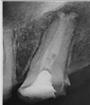
The prevalence of fused roots and consequent merged and C-shaped canals in maxillary second molars can be unpredictable. Thus, pose a clinical challenge. The purpose of this article was to present 2 cases with a C- shaped configuration diagnosed during root canal treatment. Both Cases reflect the fusion into a C-shaped configuration and the configuration classified as Type B2. Clinicians should be aware of possible anatomic variation of the root canal system and anticipate such complexities when formulating an effective root canal treatment plan. Moreover, when a preoperative periapical radiograph shows signs of a fused-rooted maxillary second molar, utilization of operative dental microscopes coupled with Cone-beam computed tomography (CBCT) images is essential for localization and identification of all canals.
Downloads
Article Metrics Graph
##plugins.themes.bootstrap3.article.sidebar##
##plugins.themes.bootstrap3.article.details##

This work is licensed under a Creative Commons Attribution 4.0 International License.