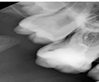
Maxillary First Molar with Seven Root Canals: A Case Report
##plugins.themes.bootstrap3.article.main##
Keywords
Abstract
The maxillary first molar exhibit unpredictable root canal morphology and different number of root canals has been reported in the literature. Thus, precise knowledge of the root canal configuration and its variation is important during root canal treatment. Moreover, detection and proper disinfection of all canals is essential to yield favorable therapy outcome. The purpose of this article is to report a case of a young Saudi female with an anatomical variation. The patient underwent Cone-beam computed tomography (CBCT) examination, and CBCT scan slices revealed seven canals: three mesiobuccal (MB1, MB2, and MB3), two distobuccal (DB1 and DB2), and two palatal (P1 and P2). All canals were successfully cleaned and filled with a single cone obturation. Recognition of such anatomic variation can be challenging. Nevertheless, CBCT examination is an excellent tool for identifying and managing complex root canal systems. In addition, utilization of dental operating microscopes can be also helpful.


